What Kind of Pain Care Would JFK Get Today?

By Pat Anson, PNN Editor

This year marks the 60th anniversary of the assassination of President John F. Kennedy, an event that shocked the world. Kennedy was only 46 when he died in 1963.

At the time, Kennedy was widely seen as a healthy, handsome and vigorous man. The truth, which emerged years later, is that JFK was chronically ill almost from birth. Scarlet fever nearly killed him as an infant, and as a child he was thin, sickly, and suffered from chronic infections and digestive problems.

Not until decades after his death did we learn that Kennedy was born with an autoimmune condition called polyglandular syndrome, and that a series of failed back surgeries may have led to adhesive arachnoiditis, a chronic and painful inflammation in his spinal canal. Historians and physicians also confirmed rumors that JFK suffered from Addison’s Disease, a well-guarded family secret.

Kennedy was given the last rites at least twice before becoming president and reportedly told his father that he would “rather be dead” than spend the rest of his life on crutches, paralyzed by pain.

In short, it’s a bit of miracle that JFK even lived to see his 46th birthday. The American public never had a full understanding of his health problems until long after he was dead.

How did Kennedy pull it off? The answers can be found in Dr. Forest Tennant’s latest book, “The Strange Medical Saga of John F. Kennedy.”

“The reason I decided to write it was mainly that I had become aware that he was an intractable pain patient,” says Tennant, a retired physician and one the world’s foremost experts on arachnoiditis and intractable pain. “Fundamentally, my book is really taking a lot of other people's work and putting it together in a historical chronological fashion. I just felt it needs to be done to really understand what happened to him.”

Although Kennedy’s chronic health problems were largely hidden from the public, many of his medical records still exist – a reflection of his family’s wealth and access to the best medical care available. Good doctors keep good medical records, especially when their patients are rich and famous.

“Until I got into doing this, it was not appreciated by me. Unless a person is very famous and has a lot of medical records, physicians never get to see a case from start to finish. Meaning from birth to death. I've never really realized how rare that is,” says Tennant.

A Controversial Drug Cocktail

In the mid 1950’s, Kennedy found a team of innovative medical experts who helped relieve his pain, elect him as president, and achieve his best health ever while living in the White House.

Dr. Max Jacobson put Kennedy on a controversial “performance enhancing” cocktail. The ingredients were secret, but Tennant says the cocktail probably included methamphetamine, hormones, vitamins and steroids.

Exhausted from months of campaigning, Kennedy was injected with the cocktail just hours before his first debate with Richard Nixon, a nationally televised debate that likely won the election for Kennedy because he appeared more energetic than Nixon.

Kennedy continued taking the cocktail as president, over the objections of White House physicians.

High Dose Opioids

Dr. Janet Travell also played a key role in revitalizing Kennedy’s health, putting him on a comprehensive pain management program that included physical therapy, hormone replacement, anti-inflammatory drugs, and the opioids methadone, codeine and meperidine (Demerol).

One of the first things she noticed was the callouses under Kennedy’s armpits from using crutches so often.

“On the day she met him, she put him in a hospital and started methadone that day, as a long-acting opioid, and then she also had him on Demerol and some other miscellaneous opioids. But his two main opioids were methadone and then Demerol for breakthrough pain,” said Tennant.

The precise dosage given to JFK is unknown, but Tennant estimates it was initially 300 to 500 morphine milligram equivalents (MME) a day, a level that would be considered risky under the CDC’s 2016 opioid prescribing guideline. The guideline recommended that dosages not exceed 90 MME.

“It would have exceeded the CDC guidelines by far,” says Tennant. “The methadone dose would have exceeded the CDC guidelines itself. But she knew to put him on methadone and if it hadn't been for methadone, he’d never have been president. He had to have something to stabilize himself right at that time. And he had to have a second opioid for breakthrough pain.”

Dosages that high today would likely attract the attention of the Drug Enforcement Administration, which investigates and prosecutes doctors for writing high-dose prescriptions. Tennant himself came under scrutiny from the DEA for giving intractable pain patients high doses, and his office and home were raided by DEA agents in 2017. Tennant was never charged with a crime, but he retired from clinical practice a few months later.

Raids like that have had a chilling effect on doctors nationwide. Many now refuse to see pain patients on opioids, regardless of the dose.

“JFK would not have been welcome today in pain clinics,” says Tennant. “My patients were very similar to JFK, almost same disease, same kind of doses, and the same kind of therapies. And of course, today that is taboo. But that was the standard in the 1950’s.  

“It's only been in the last few years that the government has decided that the standard treatment that has been there for half a century is now almost a crime.”

Dr. Travell never came under that kind of scrutiny, but Dr. Jacobsen did. Dubbed “Dr. Feelgood” by critics for his unconventional treatments, Jacobson’s medical license was suspended in New York state a few years after Kennedy’s death. The 1972 Controlled Substances Act ensured that his cocktail would never be prescribed again.

Tennant says there is no evidence the cocktail harmed or impaired JFK, who was never hospitalized or bed-bound during the 8 years he was under Jacobson’s care.

Without Jacobson and Travell, Tennant believes it unlikely Kennedy would have run for president or been elected.

“That's one of the reasons why I wrote the book. I think people need to know that JFK’s treatment was opioids. And his treatment was the standard of the day, up until the recent fiat by the federal government and state medical boards. The country got along for half a century with those standards quite well,” Tennant said.

In addition to his book about JFK, Tennant has written books about Howard Hughes and Elvis Presley, who also lived with -- and overcame -- chronic health problems.

The Tennant Foundation gives financial support to Pain News Network and sponsors PNN’s Patient Resources section.  

How Realistic Is a Possible Treatment for Hypermobile EDS?

By Crystal Lindell, PNN Columnist

Ever since I was diagnosed with hypermobile Ehlers-Danlos Syndrome (hEDS) back in 2018, I’ve spent countless hours contemplating what my life would be like if there was an effective treatment.

Last week, Tulane University School of Medicine pushed that question to the forefront. They announced that they may have found a potential cause of hEDS and hypermobility in general. They also theorized a potential treatment.

Ehlers-Danlos Syndrome (EDS) creates fragile connective tissue, which can cause a range of other symptoms including joint pain, digestive trouble, migraines and more.  

Tulane researchers proposed that a variation of the MTHFR gene that causes a deficiency of folate – the natural form of vitamin B9 – could hold the key to hypermobility and a range of connective tissue disorders such as EDS. As for treatment, they hypothesized that methylated folate — which is folate that is already processed — could be a possible treatment for the myofascial pain that’s common for hypermobile patients. They published these preliminary findings in the journal Heliyon.

First, it should be noted that none of this was based on a clinical study of patients. Rather, it’s a proposal that they believe should be researched further. As such, there’s been some valid criticism of Tulane’s announcement from the EDS community. Was it premature? Will any of this hold up in a peer-reviewed study? Or are they just giving false hope to the thousands of EDS patients desperate for some good news?

As an EDS patient myself, I understand those concerns and I very much sympathize with them. Many of us have spent too much time being dismissed by doctors, and too much money on treatments that don’t end up working.

Unfortunately, I think we’re going to have to live in the uncertainty right now. We won’t know if this is a viable option until we get a peer-reviewed study. But I am glad that they are looking into this and sharing these types of updates as the research progresses. I’d rather be informed along the way than only be told at the end of the process.

What Would a Treatment Mean for Patients?

What if their theory is correct though? Is that a good thing? I have to confess, my feelings about it are complicated.

The thing about EDS is that it’s different for everyone. As it stands, most doctors treat the symptoms, which, as mentioned above, vary widely. For me, the most debilitating one is intercostal neuralgia — which is not a type of myofascial pain. As such, it seems unlikely that methylated folate would do much to treat it.

But hey, maybe methylated folate is more of a preventative treatment? That alone would be an incredible advancement for hEDS patients.

The question is, would this treatment impact anything else? Do they know if it would only address myofascial pain? Or could it also help with other symptoms? And would that even be a good thing if it did?

EDS touches every aspect of my body and even most aspects of my personality. There’s the velvety skin everyone comments on when they shake my hand. And the constant comments about how I look for my age, which also seems to be related to how EDS impacts my skin. Would methylated folate treatment change that at all? How so? And what would I end up looking like?

The most well-known EDS symptom is probably loose joints, which means they easily overextend. For me, that’s meant a lifetime of sprained ankles and joint pain. But again, what happens if methylated folate changes that? My joints may be loose, but they’re the only ones I’ve ever known. Would stiffening them up actually help me at this point? Or would I have to re-learn how to move my own body?

What about my mind? EDS patients have higher than average rates of neurodivergence like autism and ADHD. They also have higher rates of mental health issues like depression and anxiety. Could this treatment change our brains too? Or, if not, what if they eventually find a treatment that does? Would it alter aspects of people’s personalities?

There’s also the more logistical issue of EDS as a name at all. According to the National Library of Medicine, a syndrome refers to a group of symptoms and physical findings without a direct cause. Once a cause is found, the symptoms are typically renamed as a “disease.” So, if the researchers at Tulane did find a cause, what do we have? Ehlers-Danlos disease? EDD?

One thing we do know is that EDS and hypermobility cause immeasurable pain and suffering for a lot of people, so we do need more research into potential treatments. Time will tell if folate treatment proves effective or not, but either way, I hope that there are a lot more potential treatments coming.

Crystal Lindell is a freelance writer who lives in Illinois. After five years of unexplained rib pain, Crystal was finally diagnosed with hypermobile Ehlers-Danlos syndrome. She and her fiancé have 3 cats: Princess Dee, Basil, and Goose. She enjoys the Marvel Cinematic Universe, Taylor Swift Easter eggs, and playing the daily word game Semantle. 

Researchers Find Possible Cause of Hypermobile EDS

By Crystal Lindell, PNN Columnist

Do we finally have a genetic link for hypermobile Ehlers-Danlos syndrome?

Researchers at Tulane University School of Medicine think so – and it could even point to an effective treatment that’s already available.

A variation of the MTHFR gene that causes a deficiency of folate – the natural form of vitamin B9 – could hold the key to hypermobility and a range of associated connective tissue disorders such as Ehlers-Danlos (EDS), according to preliminary findings published in the journal Heliyon.

“You’ve got millions of people that likely have this, and until now, there’s been no known cause we’ve known to treat,” said Gregory Bix, MD, director of the Tulane University Clinical Neuroscience Research Center. “It’s a big deal.”

People with the genetic variant can’t metabolize folate, which causes unmetabolized folate to accumulate in the bloodstream. The resulting folate deficiency in other parts of the body prevents key proteins from binding collagen to the extracellular matrix, which plays an important role in cell growth. This leads to more elastic connective tissue, hypermobility, and a potential cascade of associated conditions, researchers said.

The discovery could help doctors more accurately diagnose hypermobility and hypermobile EDS by looking for elevated folate levels in blood tests and the MTHFR genetic variant.

“Hypermobility is widespread and unfortunately under-recognized,” said Jacques Courseault, MD, medical director of the Tulane Fascia Institute and Treatment Center. “I’m excited about being able to treat the masses where people aren’t going their whole lives being frustrated and not getting the treatment they need."

Doctors discovered the connection between folate deficiency and the MTHFR gene by working with patients at Tulane’s Hypermobility and Ehlers-Danlos Clinic, the only clinic in the U.S. that focuses on fascia disorders. Blood tests of hypermobile patients revealed elevated levels of unmetabolized folate. Subsequent tests showed that most of those with elevated folate serum levels had the MTHFR genetic variant.

The good news is a treatment already exists. Methylated folate – folate that is already processed – is FDA-approved and widely available.

“It’s an innocuous treatment,” Bix said. “It’s not dangerous, and it’s a vitamin that can improve people’s lives. That’s the biggest thing: We know what’s going on here, and we can treat it.”

We’ve discovered something in medicine that can help, not a small group of people, but potentially many across the world.
— Dr. Jacques Courseault

Though more studies and clinical testing needs to be done, researchers say patients who have been treated with folate have shown improvement: less pain, less brain fog, fewer allergies and improved gastrointestinal function.

“We’ve discovered something in medicine that can help, not a small group of people, but potentially many across the world,” Courseault said. “This is real, it’s been vetted out well and clinically we’re noticing a difference.”

What Is Hypermobile EDS?

For those with hypermobile Ehlers-Danlos syndrome (EDS), the same conditions that create fragile connective tissue can cause a range of other symptoms that, on the surface, can seem unrelated: joint pain, chronic fatigue, thin tooth enamel, dizziness, digestive trouble and migraines, as well as psychiatric disorders such as anxiety and depression. Women with hypermobile EDS may also be at increased risk for endometriosis or uterine fibroids.

For years, researchers have struggled to find the cause of hypermobility and hypermobile EDS. Of the 13 subtypes of EDS, hypermobile EDS comprises more than 90 percent of cases. But until this study, hypermobile EDS was the only subtype without a known genetic correlate. As a result, symptoms have often been treated individually, without EDS being recognized as the likely cause.

Until now, hypermobility could only be diagnosed by the Beighton score, a somewhat controversial physical exam that involves measuring the bend of the spine, fingers and limbs. There has also been a historic lack of acceptance of hypermobility as a disorder that requires specialized treatment.

Many patients with hypermobile EDS never get a proper diagnosis. As a result, the number of people with hypermobility is unclear, though it could comprise more than half the world’s population.

“Hypermobility is not rare,” Courseault said. “Hypermobility is like a Ferrari that requires a lot of maintenance and the best synthetic oil. After knowing a patient's name and date of birth, I think it's prudent for clinicians to know which of these body types they have.”

Crystal Lindell is a freelance writer who lives in Illinois. After five years of unexplained rib pain, Crystal was finally diagnosed with hypermobile Ehlers-Danlos syndrome. She and her fiancé have 3 cats: Princess Dee, Basil, and Goose. She enjoys the Marvel Cinematic Universe, Taylor Swift Easter eggs, and playing the daily word game Semantle. 

Polypeptides: A Promising Treatment for Intractable Pain

By Dr. Forest Tennant, PNN Columnist

We have long noted that persons with adhesive arachnoiditis (AA) and other causes of intractable pain who follow a high protein diet and take amino acid supplements usually have better treatment outcomes. They need fewer opioids, function better, and have a good quality of life.

Protein is composed of about two dozen separate building blocks called amino acids. Once ingested, they chemically join together and cause specific effects in the body. When two or more amino acids combine and partner, they are called polypeptides. Two polypeptides that relieve pain and heal damaged tissue are KPV (lysine-proline-valine) and Body Protection Compound or BPC-157.

KPV is useful primarily for pain relief and to reduce inflammation in the brain and spinal canal. It activates the neurotransmitters endorphin and melanocortin, which are stored in the hypothalamus. 

BPC-157 is a chain of 15 amino acids. Its primary function is to regenerate and heal tissue, including neural tissues, receptors, arachnoid membrane, cartilage and intervertebral discs. We believe it also helps heal spinal fluid leaks. BPC has a great effect on the stomach and intestine. 

Because polypeptides (PP’s) are fundamentally a conglomeration of food particles, when swallowed they are digested in the stomach and lose much of their effectiveness. That is why KPV and BPC-157, like insulin, are often taken by subcutaneous injection. Both PP’s are also available in non-injection formulations. KPV comes in an oral or nasal spray, while BPC-157 is available as sublingual tablet taken under the tongue or as a spray. 

A list of several companies that supply PP’s online is available in this bulletin.  

Who Should Take Polypeptides  

We have long-recommended a three-component medical protocol for AA and other causes of intractable pain to (1) suppress inflammation and autoimmunity, (2) regenerate tissue and (3) relieve pain. Our starting protocol for AA is now changed and anchored with KPV and BPC-157.  

To start, we recommend daily use of a polypeptide for a week. After a week, use it 3 to 5 days a week. Some persons with AA like to use KPV daily as it greatly reduces pain. Others can become tolerant to polypeptides, so skipping some days will keep the polypeptide active and effective.  

All persons with AA and/or intractable pain should, in our opinion, try the two PP’s provided here to enhance pain relief, promote tissue regeneration and healing, and reduce the use of potent medications, including corticosteroids, ketorolac, benzodiazepines, and opioids.  We also believe PP’s can reduce the use of risky surgery and invasive procedures.  

Several other polypeptides are being studied, and the Tennant Foundation will keep you apprised of new discoveries and developments. We consider polypeptides a major advance in the treatment of AA and other intractable pain conditions. 

Forest Tennant, MD, DrPH, is retired from clinical practice but continues his research on the treatment of intractable pain and arachnoiditis. This column is adapted from bulletins recently issued by the Tennant Foundations’s Arachnoiditis Research and Education Project. Readers interested in subscribing to the bulletins should click here.

The Tennant Foundation gives financial support to Pain News Network and sponsors PNN’s Patient Resources section.  

How TMS Helped Me Feel Better Physically and Mentally

By Madora Pennington, PNN Columnist

Chronic pain is often accompanied by depression. Many clinicians used to think that pain was caused by psychological distress, so they offered patients antidepressants with the attitude that their suffering was “all in their head.”

But now it is better understood that chronic pain can cause depression. Both conditions have a similar pathology and change the brain in similar ways. That is why treatments that work on depression (like antidepressants) may reduce the brain’s sensitivity to pain.

“Regardless of the cause of the pain, anxiety and depression increase the sensation of pain. Pain increases depression and anxiety, creating a vicious cycle. Breaking that cycle can help decrease pain,” says integrative physician and pain doctor Dr. Linda Bluestein.

I have Ehlers-Danlos Syndrome (EDS). Debilitating pain has been my companion since I was 14 years old. My body makes collagen that is not structurally sound. Because I am “loosely glued together,” I get injured easily because my joints are unstable and my body has a poor sense of where it actually is in relation to itself and the outside world. My thin and stretchy connective tissue sends pain signals to my brain, even when I am not injured.

It is probably not realistic for someone with Ehlers-Danlos to expect to have a life without pain, so I welcome medical treatments that might lessen my pain, even if they don’t eliminate it. My goal is to have pain that does not incapacitate me or ruin my life by taking all my attention. Thankfully, there are modalities that do this.

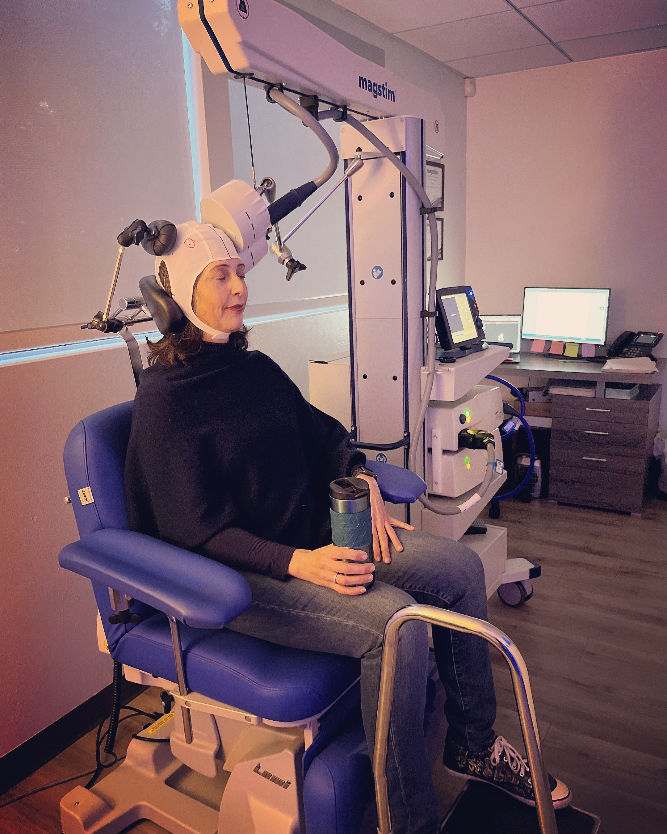
MADORA GETTING TREATMENT AT UCLA’S tms CLINIC

The last one I tried was transcranial magnetic stimulation (TMS), which stimulates the brain through a magnetic pulse which activates nerve cells and brain regions to improve mood.

TMS treatments are painless and entirely passive. The patient just sits there and lets the machine do the work. A magnetic stimulator rests against the head and pulses, which feels like tapping or gentle scratching.

TMS has been around for almost 40 years. The first TMS device was created in 1985 and the FDA approved it for major depression in 2008. Since then, its use has been expanded to include migraine, obsessive compulsive disorder, and smoking cessation.

While other medical procedures work on an injured body part, TMS targets the brain, where pain is processed. This helps the brain shift away from perceiving pain signals that are excessive and have become chronic.

“Many people are surprised to learn that stimulating the brain can help alleviate pain that is felt in an arm, leg or some other part of the body. We explain to patients that because pain is perceived in the brain, it is possible to reduce or sometimes even eliminate it by stimulating specific brain regions,” says Andrew Leucther, MD, a psychiatrist who heads UCLA’s TMS clinic, where I was treated. In addition to depression, the clinic also treats fibromyalgia, neuropathy, nerve injury, and many other causes of pain.

“Most patients are much less bothered by pain after treatment and report that they are functioning better in their work and personal lives,” Leucther told me.

Many insurers cover TMS for depression, but it is not generally covered for pain alone — although many doctors will add protocols for pain when treating depression. This is how I got my 36 sessions of TMS treatment, the usual number that insurance will cover and is thought to be effective.

Repetitive TMS stimulation to the primary motor cortex of the brain has robust support in published studies for the treatment of pain. It seems to work particularly well for migraines, peripheral neuropathic pain and fibromyalgia. Like all treatments, it may not work for everyone.

TMS practitioners recommend four or five sessions per week, gradually tapering off toward the end. My body is so sensitive, about three per week was all I could tolerate comfortably. The appointments lasted a brief 10 -15 minutes. A downside of the TMS machine is that it puts pressure against the head, which could be too much for Ehlers-Danlos patients who have uncontrolled head and neck instability.

TMS gave me relief in different ways than other methods have.  One of the first things I noticed was less negativity and rumination. It was like getting a nagging, negative person out of the room -- or rather, my head. I felt less heartbroken over the major losses of my life, such as having spent so much of it totally disabled.

I also noticed a big difference in my PTSD triggers. I found myself shrugging off situations that normally would put me in a very uncomfortable, perturbed state. Keep in mind, I was getting TMS applied to various points on my scalp for pain, depression and anxiety.

Since having TMS, I notice that my body is less sensitive to touch. From spa treatments to medical procedures, it does not hurt as much to be poked at or pressed on. The extra comfort TMS had given me, both mentally and physically, is a lot for someone with medical problems like mine that are so difficult to treat.

Madora Pennington is the author of the blog LessFlexible.com about her life with Ehlers-Danlos Syndrome. She graduated from UC Berkeley with minors in Journalism and Disability Studies. 

Why Healing Is Just as Important as Relieving Pain

By Dr. Forest Tennant, PNN Columnist

We all want to take a pill and have our pain lessen or go away, even for a moment. That is human nature. However, we must also take steps to promote healing of our damaged tissues, which over time will have a more lasting effect in reducing the severity of pain. 

There is a difference between symptomatic and healing treatment. Both are necessary to have an effective treatment program. Symptomatic treatment only relieves pain temporarily.  

We regularly hear from persons with adhesive arachnoiditis (AA) who can’t understand why their disease is progressing and why they are deteriorating. The stimulus for this topic was a man with AA who has an implanted morphine pump and an implanted electrical stimulator in the calf of his leg. He takes 15 mg of oral morphine three times a day, as well as gabapentin (Neurontin). 

He did not use a single “healing measure” and had gained so much weight he was now a diabetic. Despite his treatment, which carried a price tag of about a quarter of a million dollars, he wondered why he was deteriorating.

Every disease with the moniker “itis” — including arachnoiditis — is caused by an inflammatory and/or autoimmune process.  This simply means that your painful, damaged tissue is under constant attack. You must either diligently and persistently fight back – every day -- with healing measures or you will deteriorate and die before your time.

Examples of Symptomatic Treatment 

  • Analgesics: opioids, benzodiazepines, gabapentin, pregabalin, antidepressants

  • Implanted Stimulators

  • Implanted Pumps

Examples of Healing Measures

  • Protein

  • Walking

  • Water Soaking

  • Weightlifting

  • Oxygenation

  • Stretching

  • Amino Acids/Peptides

  • Collagen

  • Hormones

  • Vitamins

  • Anti-inflammatories

  • Electromagnetics

It is human nature to desire fast, immediate relief from pain. You must, however, start healing measures at the same time you begin symptomatic pain relief, so damaged tissues won’t deteriorate further and pain won’t increase.

A major problem is misleading advertising of expensive treatments such as implanted electrical stimulators and pain relieving drugs that lead a person into thinking the treatment has healing properties when it only provides temporary, symptomatic pain relief.

That’s why it is so important to follow our 3-component medical treatment protocol to relieve pain, suppress inflammation and autoimmunity, and heal damaged tissue.

Forest Tennant, MD, DrPH, is retired from clinical practice but continues his research on the treatment of intractable pain and arachnoiditis. This column is adapted from bulletins recently issued by the Tennant Foundations’s Arachnoiditis Research and Education Project. Readers interested in subscribing to the bulletins should click here.

The Tennant Foundation gives financial support to Pain News Network and sponsors PNN’s Patient Resources section.  

Stress Can Cause Epstein Barr Virus to Reactivate   

By Dr. Forest Tennant, PNN Columnist

More than one medical professional and a lot of persons with adhesive arachnoiditis (AA) have asked why we have been studying the Epstein Barr Virus (EBV) and its relationship to AA.  After all, almost everyone over age 25 will show EBV antibodies on a blood test. 

Our on-going studies of persons with AA continue to show that essentially all have elevated EBV antibody levels. In addition, AA almost always follows a cascade of conditions known to be associated with autoimmunity. These include fibromyalgia, chronic fatigue syndrome, neuropathies, burning feet or mouth, irritable bowel, Tarlov cyst, thyroiditis, spinal fluid leaks and deteriorated bulging discs. This cascade also appears common in persons with Ehlers-Danlos type syndromes.

Two recent comprehensive reviews on EBV support the findings in our studies. One is “Epstein Barr Virus and Neurological Diseases” by Nan Zhang, et al, and the other is “Epstein Barr Virus (EBV) Reactivation and Therapeutic Inhibitors” by Jonathan Kerr.

Both reports state that EBV infection occurs in 95% or more of people, as the human lymphocyte is its natural, lifelong habitat. Lymphocytes are white blood cells that help our immune systems fight cancer, viruses and bacteria. EBV infections are spread by saliva or other body secretions, and the initial infection usually occurs before age 25, causing a cold, sinusitis, bronchitis or infectious mononucleosis.

Once the initial infection subsides, the virus imbeds itself in lymphocytes and remain there for life. As a result, virtually all humans carry low levels of EBV antibodies. Adults with AA don’t always show a positive test for the initial EBV infection, but they do show high levels of antibodies -- meaning they carry EBV in their lymphocytes that can multiply, reproduce and reactivate at any time.

Kerr reviewed research on medicinal agents that may inhibit this reactivation, which most likely occurs during periods of stress. Our standard 3-component medical protocol contains some of the agents that Kerr believes may be effective in suppressing reactivation. These agents include acyclovir, cimetidine, vitamins A, C, and D, resveratrol, luteolin, apigenin, curcumin, astragalus, L-arginine, delta-9-tetrahydrocannabinol, and green tea.

EBV-infected lymphocytes can cross the blood brain barrier to produce neuroinflammation and tissue deterioration. EBV produces antibodies that contain biologic elements that can produce on-going autoimmune and neurological diseases. The Zhang report states that high levels of EBV antibodies “can be biologic markers that assess the risk of developing” neurologic diseases.

On-going EBV autoimmunity is indicated by high antibody levels. Reactivation of EBV may accelerate inflammation and tissue deterioration. Our longstanding 3-component medical protocol seems to help suppress both the on-going autoimmunity and the reactivation of EBV.

EBV remains harmless and dormant unless our natural resistance becomes deficient, either due to a genetic disease such as Ehlers-Danlos Syndrome or a stressful event that lowers cortisol and raises adrenalin, such as trauma, infection and psychological issues. Medical procedures such as epidural injections, spinal taps, and surgery are stressful and may also reactivate EBV. All persons with AA should determine their EBV autoimmune status.

Forest Tennant, MD, DrPH, is retired from clinical practice but continues his research on the treatment of intractable pain and arachnoiditis. This column is adapted from bulletins recently issued by the Tennant Foundations’s Arachnoiditis Research and Education Project. Readers interested in subscribing to the bulletins should click here.

The Tennant Foundation gives financial support to Pain News Network and sponsors PNN’s Patient Resources section.  
4 Oldies But Goodies That Relieve Back Pain

By Dr. Forest Tennant, PNN Columnist 

In our studies, we routinely review persons with adhesive arachnoiditis (AA), Ehlers-Danlos Syndrome, Tarlov cysts and Epstein Barr autoimmunity. Our bulletins have, for the last two years, focused on new discoveries such as autoimmunity, medicinal agents, spinal fluid flow exercises, MRIs and laboratory testing.  

It’s a little embarrassing, but we haven’t sufficiently emphasized that some older treatments can still bring a lot of relief.  

AA causes considerable imbalance and unusual stretches to the muscles, tendons, nerves, and joints in the back, hips and pelvis. Consequently, these tissues become sprained, strained and inflamed. Many “old-time” measures can heal these tissues and enhance comfort and mobility. Here is a short summary of four that will be around for a long time since they simply provide comfort and relief. 

  1. Ultrasound: Several years ago, we started using ultrasound for AA. The theory is that it may break up adhesions. While this may or may not happen, ultrasound often provides immediate relief that can last days or weeks. Medications such as cortisone cream can also be administered during ultrasound, which boosts their effectiveness. There are now hand-held ultrasound devices that can be purchased for use at home. 

  2. Epsom Salts: Foot baths with minerals are convenient and soothing. Epsom salt baths are generally believed to “pull out” or detoxify the body of excess electricity and toxins. They can be most helpful for burning feet sensations and stabbing pains in the legs. 

  3. Heating Pad: Heat dilates blood vessels, which brings more oxygen to the treated area and promotes healing. Heat also relaxes muscles that may be in spasm. 

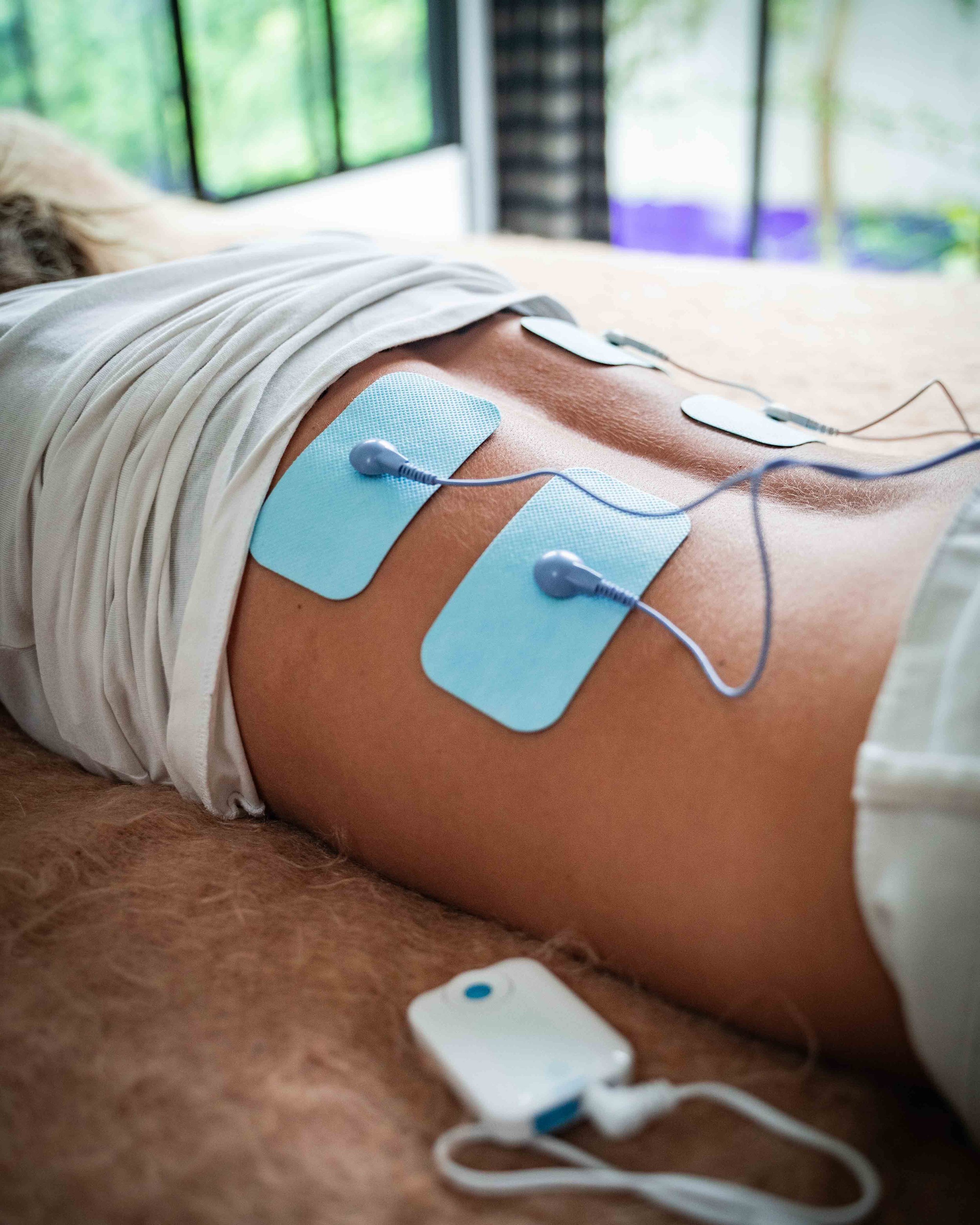
  4. Transcutaneous Electrical Nerve Stimulation (TENS): Electrical currents act as an anesthetic on nerves and nerve roots. Pain in persons with AA may temporarily abate when an electric current is administered over the lower back. TENS can often break a flare.

The human body has remained unchanged for thousands of years. Remedies and treatments discovered long ago may still be applicable today. AA has many associated conditions including spinal fluid leaks, inflammation in tissues around the spine, muscle spasm, and radiating pain among others. Some “old-time” treatments may be a welcome adjunct to the 3-component medical protocols.

Forest Tennant, MD, DrPH, is retired from clinical practice but continues his research on the treatment of intractable pain and arachnoiditis. This column is adapted from an updated bulletin recently issued by the Arachnoiditis Research and Education Project. Readers interested in subscribing to the bulletins should click here.

The Tennant Foundation gives financial support to Pain News Network and sponsors PNN’s Patient Resources section.  
Deaths of Intractable Pain Patients Often Mistaken as Overdoses

By Dr. Forest Tennant, PNN Columnist

Unexpected and sudden death commonly occur in persons who have poorly controlled pain caused by Intractable Pain Syndrome (IPS). Many persons with IPS who have died unexpectedly have been falsely accused of an “overdose” because drugs were found in their body fluids at autopsy. In reality, the cause was almost always cardiac arrest, hypoglycemia or adrenal failure.

Cardiac Arrest

Pain flares during cardiac arrest may cause the adrenal glands to pump out so much adrenalin that their blood pressure and pulse rate jump up dramatically. This causes blood vessels, including the coronary arteries and those in the brain, to constrict and shut off blood flow. The result may be a heart attack, stroke or arrhythmia.

Chronic, recurrent coronary constriction may cause heart pain called “angina.” A person with IPS who has their medications, usually opioids or benzodiazepines, reduced too rapidly is very prone to cardiac arrhythmia and cardiac arrest.

Hypoglycemia

Insulin is normally made and secreted by the pancreas to lower blood sugar in order to digest food and stabilize metabolism. In times of pain, cortisol and blood sugar are raised. When this occurs, insulin is pumped out by the pancreas to heal injured or damaged tissues. Too much insulin caused by a pain flare can force blood sugar to drop to such a low level – a condition known as hypoglycemia -- that death may occur.

The long-term effect of constant pain on the pancreas is an insulin deficiency, so high blood sugar levels (diabetes) are regularly found in persons with IPS.

Adrenal Failure

Uncontrolled constant pain may exhaust the adrenal glands to a point that the hormone cortisol drops too low, causing Addison’s disease or adrenal insufficiency. Symptoms such as darkened skin, abdominal pain and weakness usually appear slowly, but if there’s rapid onset of symptoms it could lead to adrenal failure and death.

Addison’s Disease is named after Dr. Thomas Addison, who described 11 cases of adrenal failure in 1855. About half his cases had histories of severe pain. Persons who die of adrenal failure often do so in their sleep.

Many persons with IPS have unexpectedly and suddenly died and have been falsely accused of drug overdose. To prevent sudden death, persons with IPS must be in a pain treatment program that is balanced and doesn’t rely just on just one or two medications.

Forest Tennant, MD, DrPH, is retired from clinical practice but continues his research on the treatment of intractable pain and arachnoiditis. This column is adapted from a bulletin recently issued by the Intractable Pain Syndrome Research and Education Project.

The Tennant Foundation gives financial support to Pain News Network and sponsors PNN’s Patient Resources section.   
Epstein-Barr Virus Linked to Autoimmune Conditions

By Dr. Forest Tennant, PNN Columnist 

The Epstein-Barr virus (EBV) is a herpes virus that normally resides after infection as an organism in the epithelial tissues of the throat and lymphocytes. Most humans carry the virus and blood tests will often show low levels of EBV antibodies.  

Recent research has determined that in some people, for unknown reasons, the virus will reactivate and/or produce antibodies that carry toxic elements to tissues in the body such as the spinal cord. EBV infected lymphocytes can then cross the blood brain barrier and enter the brain, spinal cord and spinal fluid.  

This situation is now referred to as “EBV autoimmunity” and is reported by multiple medical institutions and researchers to be a major, causative factor in multiple sclerosis, systemic lupus, rheumatoid arthritis, and about 2% of the world’s cancer cases.  

Autoimmunity and Arachnoiditis 

Adhesive Arachnoiditis (AA) has long been known to be an inflammatory disease in which cauda equina nerve roots become adhered by adhesions to the arachnoid lining of the spinal canal. We have also long suspected that autoimmunity was a factor in AA, but until now there has been no compelling reason for this belief. 

In our review of over 800 confirmed cases of AA by magnetic resonance imaging (MRI), along with medical history and symptoms, a single fact emerged. Almost all cases had multiple herniated or protruding intervertebral discs prior to the development of AA. These discs were often in both the cervical and lumbar-sacral regions of the spine.  

Epidural injections, spinal taps or surgery often appeared to accelerate the development of AA. But further research revealed that most persons with MRI-documented AA had other medical conditions known to be common in persons with autoimmune disease. These included: burning mouth or feet, small fiber neuropathies, fibromyalgia, carpal tunnel, Hashimoto’s thyroiditis, Sjogren’s (dry eyes), Raynaud’s, irritable bowel, migraine, temporal mandibular joint pain (TMJ), chronic fatigue, arthritis, Tarlov cysts, mast cell conditions, and POTS. Persons with a genetic connective tissue disease of the Ehlers-Danlos Syndrome type were also significantly affected.  

From this we concluded that AA is usually a late-stage component of a multisystem, autoimmune, inflammatory disease. 

Between our realization that AA is associated with multiple medical conditions and the discovery that EBV causes significant autoimmunity, we began EBV testing in persons with MRI-documented AA. Essentially every case showed very high (sometimes above laboratory testing ability) antibody levels. Some showed evidence of EBV reactivation. Another finding has been that some persons with AA have high levels of cytomegalovirus, other strains of herpes, and/or Lyme. 

EBV is now known to cause a multitude of autoimmune conditions. Our studies indicate that AA is a late-stage development of an autoimmune disorder at least partially caused by EBV. This discovery leaves us little option but to recommend that each person with AA determines if they have multiple autoimmune manifestations including herniated discs and, if so, seek EBV antibody testing and become knowledgeable about control measures. 

Forest Tennant, MD, DrPH, is retired from clinical practice but continues his research on the treatment of intractable pain and arachnoiditis. This column is adapted from an updated bulletin recently issued by the Arachnoiditis Research and Education Project. Readers interested in subscribing to the bulletins should click here.

The Tennant Foundation gives financial support to Pain News Network and sponsors PNN’s Patient Resources section.  

Why Troches Make Medications More Effective

By Dr. Forest Tennant, PNN Columnist

We regularly get emails from people who have gastrointestinal problems or tell us a particular medication is ineffective for them.  Pills and other oral medications are not always the best way to treat Adhesive Arachnoiditis (AA) or its related diseases.

The problem is that the stomach, intestine and liver don’t assimilate and metabolize more than 30 to 50 percent of the swallowed oral dose. Oral medications may also cause gastric irritation or even bleeding, and the drug may require an hour or more to be effective. Persons with AA and Ehlers-Danlos Syndrome (EDS) may have gastrointestinal dysfunction, which can make some oral medications like opioids almost totally ineffective.

If you are experiencing gastrointestinal problems or believe some of your medications are ineffective, we suggest you try using troches (the Greek pronunciation is “tro-key”). Troches are essentially lozenges that contain medication. They are placed in the mouth between the tongue and cheek until the medication dissolves.

The ancient Egyptians made some of the first troches from honey, herbs and spices to treat sore throats. Medicines introduced into the body this way bypass the digestive system and deliver their active ingredients directly into the blood stream through blood vessels under the tongue and in the cheek.

Superior Traits of Troches

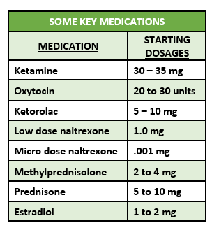
Troches are a superior way to administer many of the key medications and hormones required to treat AA, and its related problems of Tarlov cysts and EDS. Some of these medications, like ketamine and oxytocin, are essentially ineffective if swallowed. 

  • More potent than swallowed medication 

  • Fast acting – within 10 minutes 

  • No direct gastrointestinal irritation 

  • Can reduce reliance on opioids 

  • Avoids injections and suppositories

A troche must be compounded by your local pharmacy.  Every community today has pharmacies that will make or “compound” troches. For better pain relief and control of AA, Tarlov cysts and EDS, we highly recommend that patients and medical practitioners take the advanced step in therapeutics and begin to use troches. 

Forest Tennant, MD, DrPH, is retired from clinical practice but continues his research on the treatment of intractable pain and arachnoiditis. This column is adapted from a bulletin recently issued by the Arachnoiditis Research and Education Project. Readers interested in subscribing to the bulletins should click here.

The Tennant Foundation gives financial support to Pain News Network and sponsors PNN’s Patient Resources section.   

Time to Reject the Label ‘Failed Back Surgery Syndrome’

Dr. Forest Tennant, PNN Columnist

In our research studies on Adhesive Arachnoiditis (AA), a high percentage of patients tell us that their doctors have said the cause of their pain is “Failed Back Surgery Syndrome” (FBSS).

This misnomer and poor excuse for a diagnosis has fortunately been rejected “out-of-hand” as a cause of pain by most upscale physicians. After all, FBSS is neither a disease nor a symptom. We can do better in 2022.

Some three or four decades ago, FBSS became a popular moniker used by most physicians, including this author. Why? Patients who had back pain underwent surgery, which was technically and expertly done, but the pain didn’t go away. This situation was a mystery in times past, but the label FBSS should never be used or applied in this day and age.

Today, magnetic resonance imaging (MRI) has advanced so much since its invention in the late 1980’s that painful disorders and diseases of the spine can almost always be diagnosed. There is now a technique called “contrast MRI” that allows a clear distinction to be made between spinal fluid and solid tissues, including the spinal cord, discs, nerve roots and covering of the spinal canal.

In our recent research studies, we have reviewed contrast MRIs in well over 200 persons who have been labeled as having FBSS. Every person had one or more obvious reasons to have chronic pain, despite well-done, competent surgery. For example, our reviews have found discs that have protruded, adhesive arachnoiditis, epidural fibrosis, spondylolisthesis, Tarlov Cysts, arthritic vertebra or other good, solid reasons to have pain, regardless of surgery.

Let’s be clear. Spine surgery may be necessary for a number of reasons, the most common being significant herniation of a disc or collapse of vertebrae that may dangerously compress the spinal canal. It also may be fair to say that a lot of surgery can now be avoided, since eminent danger and need for surgery is pretty clear cut on modern, contrast MRI’s.    

In addition to contrast MRIs, we now have good diagnostic tests for inflammation, autoimmunity and genetic connective tissue disorders, which are emerging as legitimate causes of severe, chronic back pain that hasn’t responded to chiropractic, physical therapy, anti-inflammatory agents and corticosteroid injections.

Put simply, the diagnostic evaluation of severe chronic back pain needs a contrast MRI and specific diagnostic tests rather than a non-descript, “wastebasket” diagnosis like FBSS.

Perhaps the worst thing about the label FBSS, is that it is easy to apply and avoids the time, money and knowledge to make a specific diagnosis. These excuses to avoid a specific diagnosis must now be rejected because we have the diagnostic and treatment measures to better the lives and health of those who suffer from severe chronic back pain.

Forest Tennant, MD, DrPH, is retired from clinical practice but continues his research on the treatment of intractable pain and arachnoiditis through the Tennant Foundation’s Arachnoiditis Research and Education Project and the Intractable Pain Syndrome Research and Education Project.

The Tennant Foundation gives financial support to Pain News Network and sponsors PNN’s Patient Resources section.   

Ketamine and Oxytocin Provide Pain Relief for Arachnoiditis

By Dr. Forest Tennant, PNN Columnist

Nearly every day we receive an email from someone who is having trouble getting enough pain medication to give them a good quality of life.  In this age of opioid restrictions, there is hope. 

In the past, opioids and gamma amino butyric acid (GABA) substitutes such as diazepam (Valium) and gabapentin (Neurontin) have been the mainstays for pain control of adhesive arachnoiditis (AA). Today, there are alternatives that can enhance your current program to give you better pain control.

Low dose naltrexone is the initial pain reliever recommended to newly diagnosed AA cases. There are two other potent pain relievers that can be used with both naltrexone and opioids to achieve better pain relief: ketamine and oxytocin. Either agent is a good opioid substitute.

Ketamine provides pain relief primarily by suppressing a nerve receptor called N-methyl-d-aspartate. It can be taken by several non-oral routes of administration: nasal, injectable, sublingual or troche (dissolvable tablet).

Oxytocin (not to be confused with oxycodone or OxyContin) is a hormone that is a natural pain reliever. It surges in a woman’s body at the time of delivery to provide pain relief. It acts by activating the endorphin (opioid) receptors and by blocking nerve impulses between the brain and spinal cord.

Every community now has one or more pharmacies that will compound or “make” formulations of ketamine or oxytocin. We favor under-the-tongue (sublingual) or buccal (cheek) formulations.

Ketamine and/or oxytocin can be taken between opioid dosages or within 5 to 10 minutes before or after an opioid dosage to make the opioid stronger and last longer.

Ketamine and oxytocin can be used separately or used as combination therapy. Starting dosages of ketamine are 10-15 mg and oxytocin 10-20 units, which are administered within 10 minutes of each other. Dosages can later be raised above the starting dose.

We find the combination of ketamine and oxytocin to provide equal or better pain relief that most prescription opioids. Neither ketamine nor oxytocin are opioids, so there is no bias or resistance to their use. Also, overdoses are essentially not known to occur with regular dosages.

Forest Tennant, MD, DrPH, is retired from clinical practice but continues his research on the treatment of intractable pain and arachnoiditis. This column is adapted from a bulletin recently issued by the Arachnoiditis Research and Education Project. Readers interested in subscribing to the bulletins should click here.

The Tennant Foundation gives financial support to Pain News Network and sponsors PNN’s Patient Resources section.   

How Intractable Pain Causes Brain Tissue Loss

By Dr. Forest Tennant, PNN Columnist

The brain not only controls pain but the endocrine, cardiovascular, metabolic, respiratory and gastrointestinal systems. Any or all of these biologic systems may malfunction if there is brain tissue loss.

Beginning in 2004, brain scan studies began to document that brain tissue loss can be caused by intractable pain. Today, almost 20 years later, this important fact appears to be either unknown or a mystery to both the public and medical professionals.

Basic science researchers have unraveled the complex process of how and why this pathological phenomenon may occur. A good understanding of how this pathology develops is critical to properly care for and treat persons who develop intractable pain whether due to a disease or an injury.

What Causes Tissue Loss?

Tissue loss anywhere in the body is caused by inflammation, autoimmunity, or loss of blood supply due to trauma or disease. The brain scan studies done since 2004 that documented brain tissue loss were not done in persons who had a stroke or head trauma, but in pain patients experiencing inflammation and autoimmunity (i.e., collagen deterioration). It turns out that both biologic mechanisms may operate to cause brain tissue loss in intractable pain patients.

In the pursuit of understanding brain tissue loss and its accompanying malfunctions, it has been discovered that the brain and spinal cord (central nervous system or CNS) contain cells called microglia. They are closely akin to the immune protective cells in the blood stream which are called a “lymphocytes.”

The microglia in the CNS lay dormant until a harmful infection, toxin or bioelectric magnetic signal enters its domain, at which time it activates to capture and encapsulate the danger or produce inflammation to destroy the offender.

If the microglia are overwhelmed by some danger, such as a painful disease that isn’t cured, it produces excess inflammation that destroys some brain tissue which can be seen on special brain scans. Some viruses such as Epstein Barr may hibernate in microglia cells and create an autoimmune response, which magnifies inflammation and brain tissue loss.

Intractable pain diseases such as adhesive arachnoiditis (AA), reflex sympathetic dystrophy (CRPS/RSD), and genetic connective tissue diseases such as Ehlers-Danlos syndrome may incessantly produce toxic tissue particles and/or bioelectromagnetic signals that perpetuate microglial inflammation, tissue loss and CNS malfunctions.

This is the reason why proper pain management must have two targets: the pain generator and CNS inflammation.

How To Know You Have Lost Brain Tissue

If your pain is constant and never totally goes away, it means you have lost some brain tissue and neurotransmitters that normally shut off pain. If you have episodes of sweating, heat or anxiety, you probably have inflammation that is flaring. Naturally, if you feel you have lost some reading, calculating or memory capacity, it possibly means you have lost some brain tissue. MRI’s may also show some fibrous scars.

Fortunately, studies show that if a painful disease or injury is cured or reduced, brain tissue can regenerate. While we can’t guarantee that brain tissue will be restored, we offer here our simple, immediate and first step recommendations using non-prescription measures.

First, do you know the name and characteristics of the disease or injury that is causing your pain? Are you engaging in specific treatments to reduce or even cure your disease, or are you simply taking symptomatic pain relief medications? 

Start at least two herbal-botanical agents that have some clinical indications that they reduce inflammation in the brain and spinal cord: serrapeptase-palmitoylethanolamide (PEA) and astragalus-curcumin-luteolin-nanokinase. You can take different agents on different days. 

Increase the amount of protein (meat, fish, poultry, eggs) in your diet. Consider a collagen supplement. Limit starches and sugars. 

Start taking these vitamins and minerals:

  • Vitamin C - 2,000mg in the AM & PM

  • Vitamin B-12, Vitamin D

  • Minerals: Magnesium and selenium

We recommend vitamins daily and minerals 3 to 5 days a week. 

The above will help you stop additional tissue loss and hopefully regenerate brain tissue.  

Forest Tennant, MD, DrPH, is retired from clinical practice but continues his research on the treatment of intractable pain and arachnoiditis. This column is adapted from bulletins recently issued by the Arachnoiditis Research and Education Project and the Intractable Pain Syndrome Research and Education Project.

The Tennant Foundation gives financial support to Pain News Network and sponsors PNN’s Patient Resources section.   

Electromedical Treatments for Arachnoiditis

By Dr. Forest Tennant, PNN Columnist  

Adhesive Arachnoiditis (AA) is an inflammatory, nerve root entrapment disease in which cauda equina nerve roots are glued by adhesions to the arachnoid-dural covering of the spinal canal. An inflamed tumor-like mass is formed inside the spinal canal that blocks spinal fluid flow, allows seepage of fluid into tissue outside the spinal canal and shuts off electrical impulses that activate the legs, feet, bladder, intestine and sex organs. Autoimmunity is produced and/or magnified by AA. 

We highly recommend a three-component protocol for AA to reduce inflammation and autoimmunity, regenerate damaged tissue and to provide pain control. Recent advances in electromedical therapies can help achieve these three goals. 

There are two basic types of electromedical devices available for AA treatment: electric current therapy (EC) and electromagnetic therapy (EM). 

Electric Current Therapy 

Almost everyone is familiar with “TENS” units, which stands for “transcutaneous electrical nerve stimulation.” These devices were the first electromedical therapies to relieve pain and promote healing.

TENS units deliver a single electric current into tissues to produce an anesthetic, pain relieving effect.  

Today, more advanced EC devices administer micro-currents and/or a combination of multiple currents with different frequencies. 

Electromagnetic Therapy 

There is a form of energy that is half electricity and half magnetism, which can be divided into wave lengths. The very shortest wave of electromagnetic energy is “atomic” and the longest is “radio.” The shortest wave used in medicine is “laser.” Other electromagnetic energy waves used for medical purposes include infrared, light and microwave. 

EC and EM devices, when placed over the lower back, deliver electric current or electromagnetic energy to the lumbar-sacral spinal canal and the spine’s surrounding tissue.

Modern devices use intermittent pulsation of electric currents or electromagnetic energy to penetrate the skin and subcutaneous tissue to reach the AA site, which is usually about 2-3 inches below the skin.  

Some devices use the label PEMF, which stands for “pulsed electromagnetic frequency.” We believe that the newer EC and EM devices can deliver electric currents or electromagnetic energy that, when pulsed, penetrate deep enough to reach the AA disease site. 

Although not totally curative, these devices usually bring about pain reduction in the 20 to 30% range. Within an individual’s financial capability, we recommend that an EC and/or EM device be used 2 to 3 times a week (not daily). EC and EM therapy are not substitutes for a medical protocol. 

EM and EC devices often produce some initial healing, but later seem to stop working. In this situation the device may have done its maximal healing. The devices can still be used periodically to prevent relapses and treat flares. 

Forest Tennant, MD, DrPH, is retired from clinical practice but continues his research on the treatment of intractable pain and arachnoiditis. This column is adapted from a bulletin recently issued by the Arachnoiditis Research and Education Project. Readers interested in subscribing to the bulletins should click here.

Dr. Tennant’s new book, "Clinical Diagnosis and Treatment of Adhesive Arachnoiditis” is available on Amazon. 

The Tennant Foundation gives financial support to Pain News Network and sponsors PNN’s Patient Resources section.